
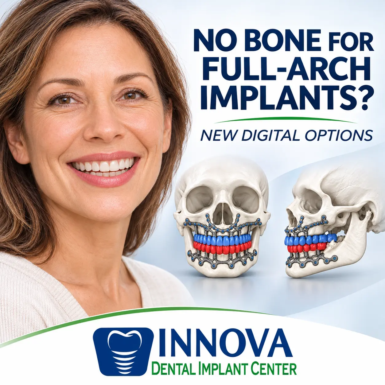
No Bone for Full-Arch Implants? New Digital Innovations
No Bone for Full-Arch Implants? Your Options & The New Innovations
Being told there is no bone for full-arch implants can feel like a closed door. Many people have worn loose dentures for years, struggled to chew, or lived with missing teeth, and then heard that the jaw is too far gone. It can sound as though the possibility of implant-supported teeth has ended.
For some patients, conventional implants are difficult or unsuitable without extensive reconstruction. Still, the field has changed. Three-dimensional imaging, digital planning, and custom titanium manufacturing have renewed interest in an older idea: patient-specific subperiosteal implants. This article explains what these options are, what they can and cannot do, and what the evidence shows so far.
A Patient Encounter That Shaped My Thinking
I recently evaluated a long-time denture wearer who had been told she did not have enough bone for conventional dental implants. Her examination and radiographs showed advanced bone loss in both jaws.
In the upper jaw, the ridge had resorbed substantially. In the lower jaw, the remaining bone was so reduced that the nerve openings sat close to the denture-bearing surface. She told me her lower denture sometimes caused tingling when it pressed on certain areas. With bone loss this advanced, pressure near the nerve may contribute to those sensations, although any symptom needs individual evaluation.
Given those findings, I understood why the earlier evaluation ruled out conventional implants. But the encounter raised a question I could not set aside: what can modern technology offer when the usual implant sites have almost no usable ridge bone?
That question led me to digitally designed subperiosteal frameworks. This article explains the concept. It does not suggest that this patient underwent the procedure or was necessarily a candidate for it.
[IMAGE PLACEHOLDER: Clinical panoramic radiograph. Before publication, confirm HIPAA-compliant de-identification or obtain the required patient authorization; have the final image and caption reviewed for patient privacy.]
Suggested caption: Advanced bone loss in both jaws can limit conventional implant placement and bring important anatomical structures close to the denture-bearing surface.
No Bone for Full-Arch Implants: What Does That Mean?
Clinicians place conventional dental implants into the jawbone. They need enough bone volume, safe positioning relative to nearby structures, and biological integration with the surrounding bone, a process called osseointegration.
After a tooth is lost, the alveolar ridge, the bone that once surrounded its root, gradually shrinks. Over the years, it may become thin or shallow. In the upper jaw, the nasal floor and maxillary sinuses can limit placement. In the lower jaw, the inferior alveolar nerve and the mental foramina create important boundaries.
As a result, some patients need extensive grafting or a different anchorage approach. However, “not enough bone for dental implants” usually refers to conventional implant sites. It does not mean there is no bone anywhere in the jaw. Other regions may remain, but not every patient’s anatomy suits an alternative system. Clinicians need a thorough examination and three-dimensional imaging to answer that.
No Bone for Full-Arch Implants? Start With the Teeth You Want
Most discussions of severe bone loss focus on surgery: where to place implants, how much bone remains, and whether grafting is needed. Those questions matter, but the patient came for teeth, not titanium hardware.
A successful plan starts with the intended restoration. We need to understand the desired tooth position, the relationship between the upper and lower jaws, the bite, lip support, speech, aesthetics, and the space available for the prosthesis. We also need to decide whether the final teeth should be fixed or removable, and how the patient will clean and maintain them.
This matters when someone hears there is no bone for full-arch implants, because the ideal tooth position may be far from the remaining bone. A framework can fit the bone precisely and still lead to a bulky, hard-to-clean prosthesis if its connections emerge in poor positions. For that reason, prosthetic planning and skeletal fixation must be developed together. Prosthodontists, surgeons, and engineers each contribute to that process.
What Is a Custom Subperiosteal Implant?
A modern subperiosteal implant is a custom metal framework that rests on selected bone surfaces beneath the gum tissue. It differs from a root-shaped implant placed into the ridge. Three components work together:
- Patient-specific titanium framework. Digital design lets the framework match the planned supporting bone. It does not need a conventional implant socket at each tooth position.
- Fixation screws. Screws anchor the framework to bone suitable for rigid support, sometimes away from the most resorbed ridge. This is mechanical fixation, not primarily the osseointegration of a root-shaped implant.
- Prosthetic pillars. Integrated posts pass through the soft tissue and connect to the restoration. Their position, height, contour, and accessibility affect the final prosthesis.
The framework is not the finished set of teeth. It is the supporting structure, and designing the restoration remains a separate, essential responsibility.
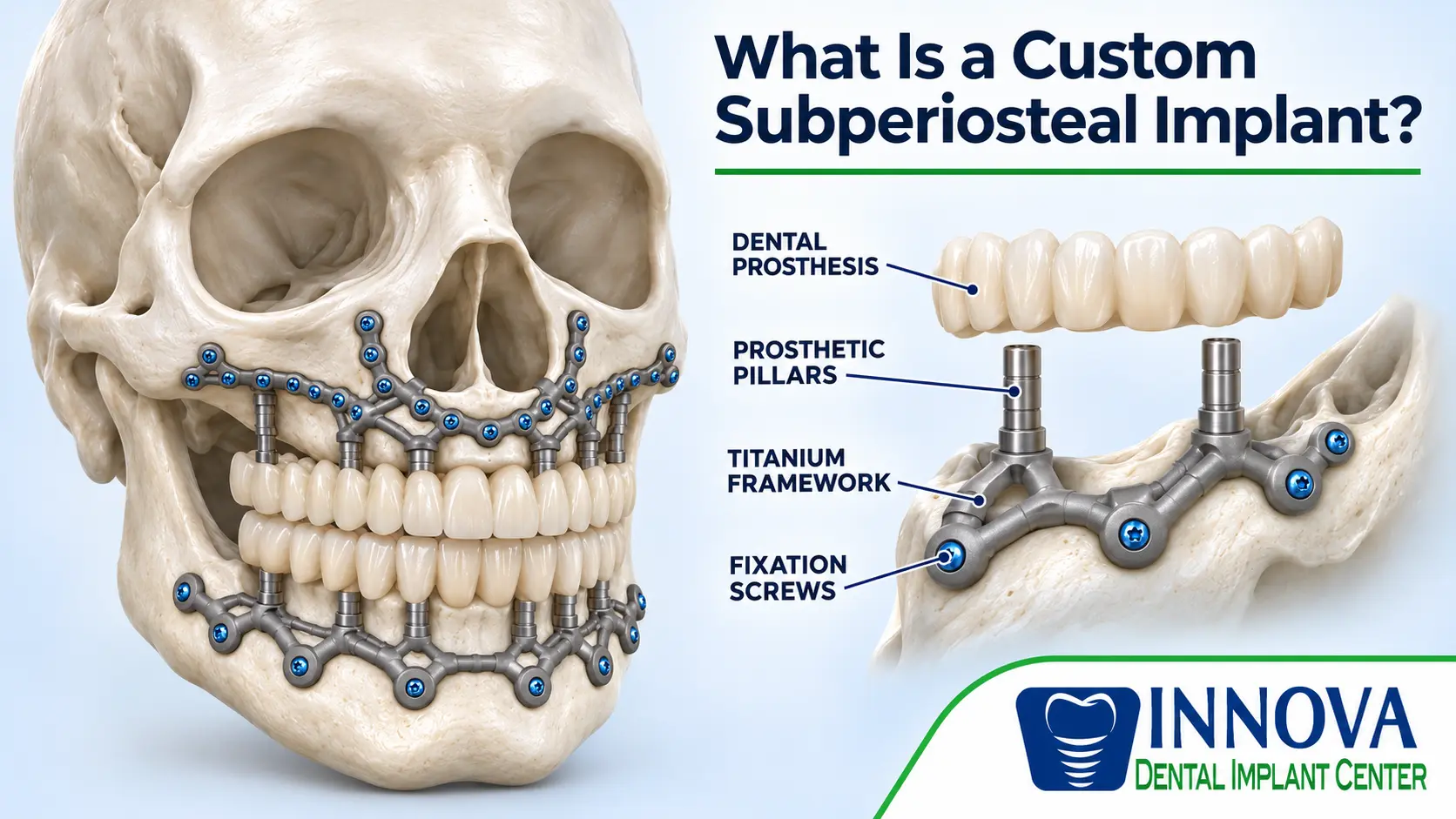
Illustration of a custom subperiosteal implant showing the titanium framework, fixation screws, prosthetic pillars, and dental prosthesis. Conceptual illustration; not an actual clinical case.
No Bone Left for Implants? An Old Idea With a New Digital Workflow
Subperiosteal implants have existed since the 1940s. Early designs often required direct impressions of exposed bone, and clinicians faced problems with fit, mobility, infection, and soft-tissue exposure. As predictable root-shaped implants became standard, these systems largely fell out of routine use.
What has changed is the workflow. CT or cone-beam CT imaging builds a three-dimensional model of the jaw. Computer-aided design helps plan the framework, its connections, and its fixation sites before surgery. Metal additive manufacturing then produces the titanium structure, often by laser melting successive layers of powder. Better manufacturing does not by itself remove surgical or biological risk, though.
Not Enough Bone for Dental Implants? How a Custom Framework Is Planned and Placed
The sequence varies by system and case, but these five steps show how anatomy, engineering, and the final teeth connect.
Step 1: Examine the Jaw in Three Dimensions
Cone-beam CT or medical CT shows bone contours, nerves, sinuses, and possible fixation areas. A panoramic X-ray gives an overview, but three-dimensional imaging shows the spatial relationships a custom framework needs. A scan alone does not determine candidacy. Tissue quality, medical history, infection risk, and surgical access also matter.
Step 2: Plan the Teeth and Their Connections
The restorative team evaluates tooth position, occlusion, vertical dimension, lip support, and space for the prosthesis, and plans hygiene access. The pillars must emerge where a workable prosthesis can attach. Otherwise the restoration may be bulky, hard to clean, or poorly positioned against the opposing teeth.
Step 3: Design and Manufacture the Framework
Clinicians and engineers refine the virtual framework to include the planned fixation sites and pillars. Manufacturers then build the titanium framework layer by layer from medical-grade titanium alloy. The computer model becomes a patient-specific support, while the restorative team designs the replacement teeth as separate components.
Step 4: Secure the Framework to Suitable Bone
During surgery, the surgeon exposes the planned bone areas, checks the fit, and fixes the framework with screws. This is still significant surgery and may require broad tissue elevation and careful protection of nearby structures. A custom framework still needs adequate bone for fixation. It cannot solve every case described as having “no bone.”
Step 5: Connect and Maintain the Prosthesis
Depending on the device and protocol, the team may attach a temporary restoration during early healing. The definitive prosthesis then needs attention to tissue health, bite, material thickness, contours, and cleanability. A stable framework does not guarantee a comfortable or trouble-free prosthesis for years.
KLS Martin IPS Preprosthetic and FDA Clearance
One example is the KLS Martin IPS Preprosthetic, a patient-specific titanium subperiosteal system with planned bone fixation and integrated prosthetic pillars. The manufacturer describes its use in severe jaw atrophy and selected complex maxillofacial defects. However, being told there is no bone for full-arch implants does not by itself establish candidacy for this system.
On July 8, 2022, the U.S. Food and Drug Administration granted the system 510(k) clearance, K210228. The stated indication covers a patient-specific titanium subperiosteal implant placed between the periosteum and supporting bone of the upper or lower jaw to support multi-unit prostheses, such as dentures. The FDA record describes digital planning, titanium-alloy additive manufacturing, fixation screws, and transgingival pillars. Read the FDA clearance and summary.
The manufacturer describes the IPS Preprosthetic as the only FDA-cleared, remotely fixated, patient-specific subperiosteal system on the U.S. market. That is the manufacturer’s own claim, and it is narrower than saying no subperiosteal device has ever been cleared. See KLS Martin’s U.S. system information and its technical workflow.
FDA 510(k) clearance is not proof of superiority or a guarantee of long-term success. Some published reports describe fixed restorations supported by modern frameworks. Readers should consider those reports alongside the cleared labeling and the limits of the evidence. A design described in a case report is not automatically an accepted standard for every patient.
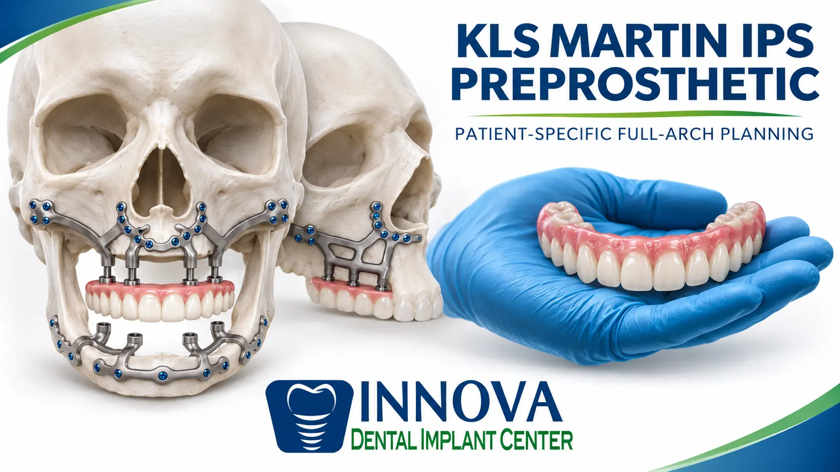
Educational illustration of patient-specific subperiosteal frameworks and a full-arch dental prosthesis. Conceptual illustration; not an official KLS Martin product rendering or actual clinical treatment result.
Can This Approach Support Fixed Teeth?
In selected cases, it may. Published clinical reports describe fixed restorations supported by patient-specific subperiosteal frameworks. The choice between fixed and removable rehabilitation depends on the device, the connection system, the anatomy, the tissue condition, and the restorative plan.
Several questions decide that choice. Can the connections support the intended teeth in the right position? Is there room for a durable restoration? Can the patient clean around the pillars and beneath the prosthesis? Can the design be maintained if repairs or adjustments are needed? These questions matter as much as the framework’s fixation.
Grafting, Zygomatic, and Pterygomaxillary Implants
Patients with severe bone loss may also hear about bone augmentation, zygomatic implants anchored in the cheekbone, or pterygomaxillary implants that use the posterior maxilla. Each has its own indications, required expertise, and risks.
Custom subperiosteal frameworks are another possible option. They are not a universal replacement for grafting, zygomatic implants, or conventional implants. The best approach depends on anatomy, health, the prosthetic goal, and the strength of the clinical evidence.
What Are the Risks, and What Does the Evidence Show?
Modern patient-specific subperiosteal implants can develop soft-tissue thinning, inflammation, infection, framework exposure, fixation problems, and prosthetic complications. Additional surgery or removal may be needed. Hygiene and follow-up matter especially when a restoration covers a complex framework.
A 2024 systematic review of additively manufactured subperiosteal implants reported that about 97.8% remained in function after a weighted mean follow-up of roughly 21 months. The same review found 25.6% had partial framework exposure. These figures combine different systems and protocols, so they are not a success rate for any single system.
More recent reviews stress that long-term evidence remains limited, particularly for soft-tissue complications and durable prosthetic function. Short-term retention should not be confused with years of comfortable, low-maintenance function. Read a 2026 review of the evidence and open questions.
For me, the key distinction is this: the framework must stay stable, and the teeth it supports must also function, remain cleanable, and be maintainable over time.
Frequently Asked Questions
Can I Get Dental Implants if I Have Almost No Jawbone?
Possibly, but not automatically. If you were told there is no bone for full-arch implants, an evaluation can show whether other reconstructive approaches are feasible. A custom framework still needs suitable bone for fixation and healthy soft tissue.
Is a 3D-Printed Framework the Same as My Final Teeth?
No. The framework provides skeletal support. A separately designed prosthesis provides the replacement teeth, and the treatment plan connects the two.
Are Custom Subperiosteal Implants Better Than Conventional Implants?
Current evidence does not establish general superiority. These systems are specialized options for selected cases where conventional implant treatment is difficult or unsuitable.
Restoring Teeth Is the Goal
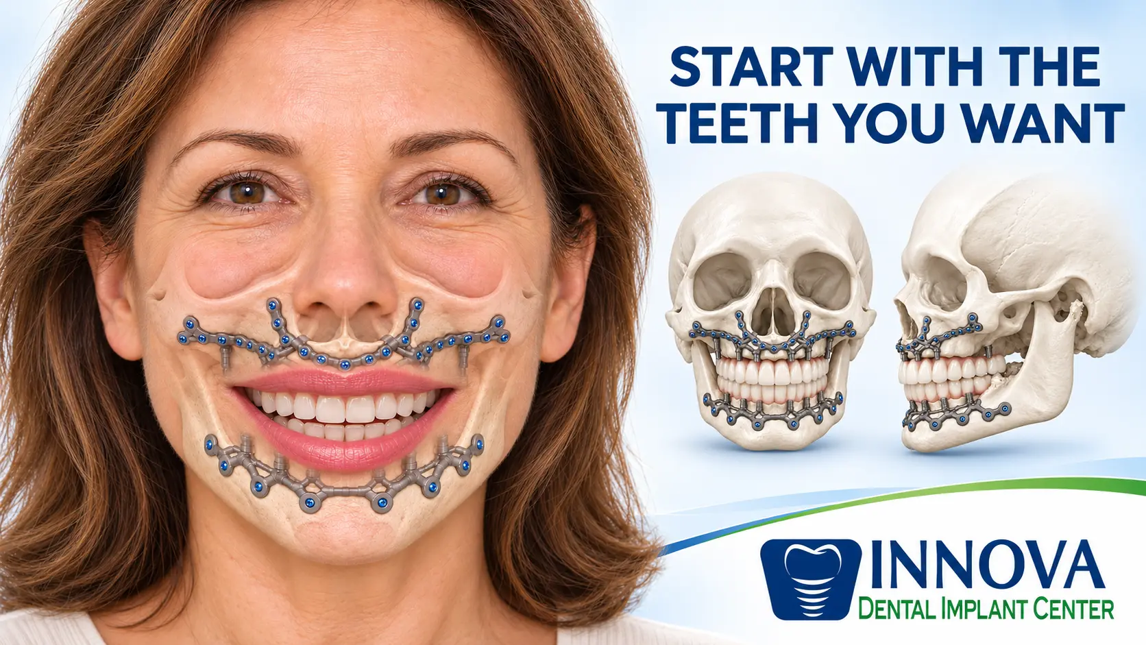
The patient I described began with a question about bone, but her reason for seeking care was more personal. She wanted to chew, speak, and smile with confidence again.
Custom subperiosteal implants show how imaging, engineering, and surgical technique can widen the discussion for selected patients with severe bone loss. Their future depends not only on better titanium frameworks but also on sound prosthetic design, healthy tissues, functional outcomes, and reliable long-term evidence.
The goal is not simply to attach titanium to bone. It is to restore teeth that serve the patient’s function, comfort, and quality of life.
About the Author
Dr. Elias Rivera, DDS, MS, FACP is a board-certified prosthodontist with advanced training in implant surgery, complex oral rehabilitation, and digital dentistry. He practices at Innova Prosthodontics & Dental Implant Center in Virginia.
Educational disclosure: This article reviews emerging technologies and published research. It is not an individual treatment recommendation. Innova Prosthodontics does not currently offer the KLS Martin IPS Preprosthetic procedure.
Scientific References
- U.S. FDA. KLS Martin IPS Preprosthetic, 510(k) K210228 (2022).
- KLS Martin. IPS Implants Preprosthetic: system and digital workflow.
- Clinical performance of additively manufactured subperiosteal implants: a systematic review (2024).
- Patient-specific subperiosteal implants for oral and maxillofacial rehabilitation: current evidence and unresolved questions (2026).
- Salvaging maxillary form and function after free tissue transfer failures: successful use of an IPS custom subperiosteal implant (2026).